
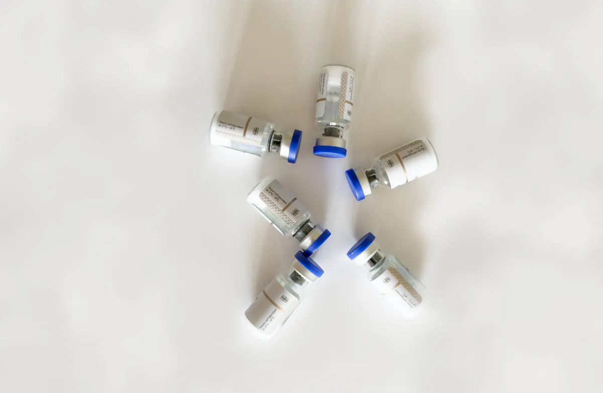
- A before and after photo documents an appearance at two moments. It does not document a cause, and it cannot separate a peptide from diet, training, lighting, posture or other products used at the same time.
- Flash alone can change how experienced clinicians judge an outcome: in one study, adding flash to one of two photographs taken seconds apart shifted surgeons' impressions of the result (Cariello et al., 2012).
- The strongest human peptide evidence sits with the prescription GLP-1 and GIP/GLP-1 medicines, where randomized trials of 1,961 and 2,539 participants measured weight change over 68 and 72 weeks.
- For BPC-157, a 2025 systematic review of orthopaedic literature retained 36 studies, of which 35 were preclinical and only 1 was clinical.
- TB-500 is a fragment of thymosin beta-4. The published human trials used full-length thymosin beta-4, applied topically to wounds or as eye drops, not injected for muscle or tendon goals.
- Cosmetic peptides have real but modest measured effects: a split-face trial of palmitoyl pentapeptide in 93 women over 12 weeks found significant fine line improvement versus the same moisturizer without the peptide.
- Visible change can coexist with harm. Case reports link melanotan use to new atypical moles and to melanoma, which is why pigment change tells you nothing reassuring about safety.
- This article is educational. It contains no dosing guidance and no protocol. Many peptides discussed are not approved for human use, and legal status varies by country.
Why do peptide before and after photos look so convincing?
Search for almost any peptide and you will find the same format: two images side by side, a few weeks or months apart, with a caption naming the compound. The format is powerful because it feels like evidence. You are not being asked to trust a statistic you cannot check, you are being shown something with your own eyes. Visual information carries an intuitive weight that a confidence interval never will.
The problem is structural rather than dishonest. A before and after pair is an observation of appearance at two moments in time. It contains no control condition, no blinding, no randomization and no measurement of anything. Even when both photos are completely genuine, the pair cannot tell you whether the peptide caused the difference, whether something else did, or whether the difference exists outside the camera at all.
There is also a selection filter operating before you ever see an image. People who perceive no change rarely photograph themselves again, and almost never post. Sellers and affiliates publish their best cases, not their median case. What reaches your screen is therefore the upper tail of a distribution whose shape you cannot see. In research terms, this is publication bias operating on social media, with no editor and no reviewer anywhere in the chain.
None of this means peptides do nothing. Several have measurable effects documented in peer-reviewed human trials, and this article walks through them. It means that the photographs and the evidence are two separate things, and that confusing them is how people end up spending money on compounds whose human data does not yet exist. The useful question is never whether a photo looks real. It is what a controlled study measured, in whom, over how long, and compared with what.
Educational content only. Nothing here is medical advice. Consult a qualified healthcare professional before considering any peptide.
What can change in a photograph while nothing changes in the body?
This is the part most readers underestimate. The variables that shape a facial or physique photograph are numerous, and several of them produce differences larger than anything a cosmetic ingredient could plausibly achieve in twelve weeks.
The clearest published demonstration concerns flash. In a study in Ophthalmic Plastic and Reconstructive Surgery, adding flash to one of two consecutive photographs taken seconds apart was enough to change how experienced surgeons judged the outcome of oculoplastic procedures. The authors concluded that this constitutes a genuine source of bias in observer-dependent studies (Cariello et al., 2012). Nothing about the patient had changed in those seconds. Only the light had.
Beyond flash, clinical photography guidance in dermatology and aesthetic practice converges on a long list of elements that must be held constant for two images to be comparable: identical lighting and background, identical camera-to-subject distance, manual rather than automatic camera settings, removal of makeup and jewellery, and consistent head position across all three axes of rotation, tilt and roll. Guidance on facial photodocumentation stresses that even minor deviations in head positioning can lead to misinterpretation of an aesthetic or surgical result.
Applied to the images circulating online, the implications are blunt. A "before" taken overhead in a bathroom under a downward ceiling light and an "after" taken at eye level near a window will differ in shadow depth around the nasolabial folds and under the eyes, regardless of the skin underneath. For physique photos, the same logic applies to pump, hydration status, tan, time of day, abdominal tension and the angle of the camera relative to the waist.
- Light direction and hardness: changes apparent wrinkle depth and skin texture
- Posture and head tilt: changes apparent jawline, jowling and eyelid position
- Makeup, moisturizer, oil or a fresh shave: changes apparent luminosity in seconds
- Camera focal length and distance: changes facial proportions and perceived volume
- Time of day and sleep: changes periorbital puffiness independently of any product
A photo pair that controls none of these is not weak evidence. It is not evidence of a causal effect at all.
What else is usually happening between the two photos?
Assume for a moment that the photographs are perfectly standardized and that the visible difference is entirely real. The attribution problem remains, because almost nobody changes one variable at a time.
People start peptides at moments of motivation. That same decision usually arrives bundled with other changes: a cleaned-up diet, a return to training, better sleep, reduced alcohol, more water, a new sunscreen habit, a retinoid, professional skin procedures, or a weight change of several kilograms. Any one of these can visibly alter skin and physique over eight to twelve weeks. Fat loss in particular changes facial contour, abdominal definition and even perceived skin tightness, which is why weight-loss photos and "skin peptide" photos often look similar.
Multi-ingredient products add a second layer of ambiguity. A serum containing a peptide typically also contains humectants, occlusives, niacinamide, antioxidants or a sunscreen filter. Hydration alone measurably improves the appearance of fine lines within hours, and consistent sun protection is the single most reliably documented intervention for photoaging. When the photo improves, the peptide gets the caption, but the formulation as a whole did the work, and the specific contribution of the peptide is unknowable without a controlled comparison.
The same applies to injected combinations. Blends and stacks are common, and when several compounds are used together with a training block and a calorie deficit, no observational photo can apportion the result. This is precisely the problem that randomization and placebo controls were invented to solve: a well-designed trial compares the peptide against the identical vehicle, in matched groups, with blinded assessors and instrumental measurement rather than impression.
Finally, there is the honest possibility of expectation effects. Self-assessed appearance and self-reported pain or recovery are subjective endpoints that respond to belief. That response is genuine for the person experiencing it, but it is not a pharmacological effect, and it does not transfer to anyone else.
Skin peptides: what do GHK-Cu, Matrixyl 3000 and collagen peptides studies actually measure?
Cosmetic and oral skin peptides have the most human data of the non-prescription categories, and that data is best described as real, instrumental and modest.
GHK-Cu (copper tripeptide). The mechanistic literature is extensive: published reviews describe GHK and its copper complex modulating matrix metalloproteinase expression and stimulating production of collagen, elastin and glycosaminoglycans in dermal fibroblasts, with broad effects on gene expression (Pickart et al., 2015; Pickart and Margolina, 2018). A 2026 systematic review of GHK-Cu and microneedle delivery screened 1,247 records and retained 64, and concluded that despite four decades of preclinical work the human clinical literature remains thin and fragmented, with most randomized trials small and based on facial cosmetic creams. Our dedicated page collects what the published work reports and what it does not: GHK-Cu before and after results.
Matrixyl 3000 and palmitoyl pentapeptide. This is one of the better-documented topical peptides. A 12-week, double-blind, placebo-controlled, split-face randomized study in 93 women aged 35 to 55 compared a moisturizer against the identical moisturizer containing palmitoyl pentapeptide (pal-KTTKS) at 3 ppm. The peptide arm showed significant improvement versus control for reduction in wrinkles and fine lines, by both quantitative image analysis and expert grading, and was well tolerated (Robinson et al., 2005). Note the comparator: the gain is over the same base cream, which is exactly the comparison a photo cannot make. See Matrixyl 3000 before and after.
Oral collagen peptides. A systematic review and meta-analysis of 19 randomized controlled trials totalling 1,341 participants found that peptides, particularly oral formulations, significantly improved skin hydration and brightness, with a modest pooled effect on wrinkle reduction, while effects on elasticity and density were inconsistent. Tolerability was good, and the authors called for larger trials with standardized outcomes. More detail on collagen peptides before and after.
The pattern across all three: instruments detect statistically significant but moderate changes in hydration, brightness and fine line parameters. That is a different claim from a dramatic visual transformation.
BPC-157 and TB-500: what is measured, and in which species?
These two are the most searched non-weight-loss peptides and the ones where the gap between online imagery and human evidence is widest. Both are frequently presented through recovery photos and anecdotes, and neither has the human trial base that such presentations imply.
BPC-157. The preclinical literature is genuinely large. Rodent studies report accelerated healing of transected Achilles tendon with in vitro stimulation of tendocyte growth (Staresinic et al., 2003), functional restoration after transected quadriceps muscle (Staresinic et al., 2006), and improved muscle healing after crush injury assessed macroscopically, microscopically, functionally and by enzyme activity (Novinscak et al., 2008). The decisive number is the species ratio. A systematic review in HSS Journal searched PubMed, Cochrane and Embase from inception to June 3, 2024 and included 36 studies: 35 preclinical and 1 clinical (Apostolakos et al., 2025). The review also notes that BPC-157 lacks US Food and Drug Administration approval and is prohibited in professional sport. A healed rat tendon is a legitimate scientific finding. It is not a human result, and it is not a photograph of a shoulder. See BPC-157 before and after.
TB-500 and thymosin beta-4. A distinction gets lost almost everywhere: TB-500 is a synthetic fragment, while the compound studied in human trials is full-length thymosin beta-4 (43 amino acids). Those trials exist, but they tested different routes for different indications. A European prospective, randomized, double-blind, placebo-controlled dose-escalation study applied thymosin beta-4 topically to venous leg ulcers across sites in Italy and Poland, with 24 patients randomized per group in a 3:1 ratio, to assess safety, tolerability and healing (Guarnera et al., 2007). A 56-day randomized, double-masked, placebo-controlled phase 2 trial of thymosin beta-4 eye drops in 9 patients with severe dry eye reported a 35.1% reduction in ocular discomfort and a 59.1% reduction in total corneal fluorescein staining versus vehicle (Sosne et al., 2015). Nine patients is a signal to investigate, not a conclusion. Neither trial studied injected TB-500 for tendon, muscle or physique outcomes. More on TB-500 before and after.
Both compounds are research chemicals in most jurisdictions, not approved for human use, and their legal status varies by country.
Why do semaglutide and tirzepatide photos have different evidentiary weight?
The GLP-1 and dual GIP/GLP-1 receptor agonists are peptides, and they are the one category where the before and after imagery is broadly consistent with large randomized human trials. The difference is that here the photos are downstream of the data rather than a substitute for it.
Semaglutide. The STEP 1 trial randomized 1,961 adults with obesity, or with overweight plus at least one weight-related condition and without diabetes, to once-weekly subcutaneous semaglutide or placebo for 68 weeks, both with lifestyle counselling. Mean change in body weight was -14.9% with semaglutide versus -2.4% with placebo, and 86.4% versus 31.5% of participants achieved at least 5% weight loss (Wilding et al., 2021).
Tirzepatide. SURMOUNT-1 enrolled 2,539 adults with obesity and without diabetes in a 72-week double-blind, randomized, placebo-controlled trial. Participants lost 16.0% to 22.5% of baseline body weight depending on the dose group (Jastreboff et al., 2022). Our page on the obesity indication is here: Zepbound before and after.
Two points matter for anyone reading photos of these medicines. First, both trials ran with structured lifestyle intervention alongside the drug, so the published figures are not drug-alone figures. Second, and most relevant to the before and after genre, the STEP 1 extension followed participants for a year after withdrawal of both semaglutide and the lifestyle intervention, and found that they regained approximately two-thirds of their prior weight loss, with cardiometabolic improvements reverting toward baseline for most variables by week 120 (Wilding et al., 2022). An "after" photo is a snapshot of one point on a trajectory, not an endpoint.
These are prescription medicines for specific indications, with documented gastrointestinal adverse effects and contraindications, and they require medical supervision. This article gives no dosing information and takes no position on whether any individual should use them. That conversation belongs with a physician.
Melanotan II: why does a visible change not mean the change is safe?
Melanotan II is the clearest illustration of why before and after photos are a poor safety instrument. Here the visible change is not subtle and not ambiguous: pigmentation darkens, and photos document it unmistakably. The dramatic imagery is precisely what makes the compound circulate on social media.
The published literature on it, however, consists largely of case reports describing dermatological harm rather than trials describing benefit. One report describes a patient who developed multiple new-onset atypical naevi within one week of receiving two melanotan injections, and the authors flagged the potential for melanotan to stimulate dysplastic naevi or possibly malignant melanoma (Reid et al., 2013). Another documents cutaneous melanoma in a context where melanocyte stimulation by melanotan-II coincided with sun bed use, noting that unlicensed use to promote pigmentation had become prevalent among young people attending fitness centres (Hjuler and Lorentzen, 2014). Further case reports describe eruptive dysplastic naevi, melanoma in situ, and mucosal changes associated with melanotan use.
Individual case reports cannot establish causation, and the authors of these papers say so explicitly. What they do establish is that the compound is biologically active on melanocytes in ways that warrant dermatological concern, that it is unlicensed, and that its sale is illegal in the United Kingdom and in many other countries. A tan is not a measure of safety. It is the visible part of a process whose less visible parts are the reason clinicians raise an alarm.
The general point generalizes beyond this one peptide: photographs capture the intended effect and almost never capture the unintended ones. Liver markers, blood pressure, mole architecture, glycaemic control and injection-site reactions do not appear in a mirror selfie. Our dedicated page covers the reports in more detail: Melanotan 2 before and after.
How strong is the human evidence, peptide by peptide?
The table below summarizes the highest level of published human evidence for each peptide discussed, and what the studies measured. It deliberately says nothing about what any individual should expect, because that is not something the published literature can answer.
| Peptide | What published studies measure | Highest level of human evidence | Dedicated page |
|---|---|---|---|
| Semaglutide | Percent body weight change over 68 weeks versus placebo, plus cardiometabolic variables | Large randomized controlled trials (1,961 participants), approved medicine | Weight loss peptides before and after |
| Tirzepatide | Percent body weight change over 72 weeks versus placebo, by dose group | Large randomized controlled trial (2,539 participants), approved medicine | Zepbound before and after |
| Collagen peptides (oral) | Instrumental skin hydration, brightness, wrinkle parameters, elasticity | Meta-analysis of 19 randomized trials (1,341 participants); modest effects | Collagen peptides before and after |
| Matrixyl 3000 / pal-KTTKS | Fine line and wrinkle reduction by image analysis and expert grading | Split-face randomized placebo-controlled trial (93 participants, 12 weeks) | Matrixyl 3000 before and after |
| GHK-Cu | Collagen and matrix gene expression, fibroblast activity, wound parameters; small cosmetic cream trials | Mechanistic and preclinical evidence extensive; human randomized trials small and fragmented | GHK-Cu before and after |
| Thymosin beta-4 (related to TB-500) | Wound closure in venous ulcers (topical); dry eye signs and symptoms (eye drops) | Small phase 2 trials of full-length Tβ4 by topical or ophthalmic route; no trials of injected TB-500 for musculoskeletal goals | TB-500 before and after |
| BPC-157 | Tendon, muscle and mucosal healing in rodent models | Systematic review retained 35 preclinical studies and 1 clinical study; no approval, prohibited in sport | BPC-157 before and after |
| Melanotan II | Case reports of atypical naevi, melanoma and mucosal changes | Case reports only; unlicensed and illegal to sell in many countries | Melanotan 2 before and after |
Read the second and third columns together. Several of these compounds have impressive biology and almost no human outcome data, which is a normal stage of drug development and an abnormal basis for personal use.
How long do the published studies actually run?
One of the most useful corrections to the before and after genre is simply looking at study duration. Online photo pairs are often captioned with intervals of two to four weeks. The trials that found measurable effects ran considerably longer.
The palmitoyl pentapeptide split-face study ran for 12 weeks. The oral collagen peptide trials pooled in the meta-analysis typically ran 8 to 12 weeks or more, with hydration changes often appearing before wrinkle parameters moved. The venous ulcer work with topical thymosin beta-4 assessed healing over roughly three months. STEP 1 ran 68 weeks and SURMOUNT-1 ran 72 weeks, with weight curves still separating from placebo well past the six-month mark.
This matters in both directions. It means that a photo taken at two weeks is very unlikely to be showing a structural change in collagen, because collagen remodelling in human skin is measured in months. It also means that short-interval photo pairs are showing something else: hydration, light, inflammation, water retention, posture, or a change in body weight.
There is a second timing issue specific to the imagery. Studies measure group averages with instruments and blinded graders. An individual in a trial can sit far from the mean in either direction, and trials also routinely document non-responders. A photo of one person at one time point tells you nothing about where in that distribution they sit, and nothing about how many people took the same compound and photographed nothing.
For a fuller treatment of the timelines reported in the literature across categories, see how long peptides take to work.
How can you read a peptide before and after photo more critically?
You will keep encountering these images, so the practical skill is triage. A short set of questions removes most of the noise without requiring any expertise in pharmacology.
- Is the lighting identical? Look at shadow direction under the nose and chin, and at specular highlights on the forehead. Different shadows mean different light, which means the pair is not comparable.
- Is the posture identical? Check head tilt, shoulder height, chin projection and camera height relative to the eyes.
- Is there makeup, oil, a tan or a fresh shave in only one image? Any of these can produce the entire visible difference.
- What else changed in that period? Diet, training, sleep, alcohol, body weight, sunscreen, retinoids, in-clinic procedures, other peptides.
- Who published it, and what do they sell? A photo attached to a product page is marketing, whether or not it is authentic.
- What is the stated interval, and is it biologically plausible? Two weeks is not enough time for dermal remodelling.
- Is there any measurement at all? Corneometry, profilometry, scale weight, imaging, blood work. A number that someone could in principle reproduce beats an impression.
The deeper shift is to stop asking peptide photos to answer a question they are not built to answer. Images are useful for documenting your own trajectory if you standardize them, which is why clinical photography protocols exist, and they are nearly useless as evidence about what a compound does in a population. For that, the trial design is the information: how many people, compared with what, measured how, over how long.
Medical disclaimer. This article is for educational purposes only and is not medical advice, a recommendation, or a protocol. It contains no dosing information by design. Several peptides discussed here, including BPC-157, TB-500 and melanotan II, are research compounds that are not approved for human use by the FDA or EMA, and their legal status varies by jurisdiction. Semaglutide and tirzepatide are prescription medicines that require medical supervision. Preclinical and animal findings do not transfer reliably to humans. Consult a qualified healthcare professional before considering any peptide, particularly if you are pregnant, breastfeeding, have a personal or family history of melanoma or other cancers, or take other medicines.
Recommended products
Research peptides selected for quality and purity:

GHK-Cu
Anti-Aging Compound
Test your knowledge
Quick quiz · 6 questions
Peptide Lab: free calculator & tracker
Calculate your reconstitution, track your peptides and injections. Free, no credit card required.
Frequently Asked Questions
Are peptide before and after photos fake?
Which peptides have the strongest human evidence behind their before and after photos?
Why do BPC-157 and TB-500 photos circulate so widely if the human evidence is thin?
Can a photo taken two weeks apart show a real skin change from a peptide?
Does an impressive before and after photo mean the peptide is safe?
Do results from before and after photos last?
If I want to document my own progress, how should I take the photos?
Is it reasonable to use a peptide based on photos alone?
Sources
- Wilding JPH, Batterham RL, Calanna S, et al. (2021). Once-Weekly Semaglutide in Adults with Overweight or Obesity (STEP 1). New England Journal of Medicine 384(11):989-1002. PMID 33567185.
- Jastreboff AM, Aronne LJ, Ahmad NN, et al. (2022). Tirzepatide Once Weekly for the Treatment of Obesity (SURMOUNT-1). New England Journal of Medicine 387(3):205-216. PMID 35658024.
- Wilding JPH, Batterham RL, Davies M, et al. (2022). Weight regain and cardiometabolic effects after withdrawal of semaglutide: The STEP 1 trial extension. Diabetes, Obesity and Metabolism 24(8):1553-1564. PMID 35441470.
- Cariello AJ, Viana GA, Osaki MH, Pamplona AL, Höfling-Lima AL (2012). Standardized clinical photography: the role of flash. Ophthalmic Plastic and Reconstructive Surgery 28(2):e41-e42. PMID 21659920.
- Robinson LR, Fitzgerald NC, Doughty DG, Dawes NC, Berge CA, Bissett DL (2005). Topical palmitoyl pentapeptide provides improvement in photoaged human facial skin. International Journal of Cosmetic Science 27(3):155-160. PMID 18492182.
- Systematic review and meta-analysis of 19 randomized controlled trials (1,341 participants) (2026). Oral and topical peptides for skin aging: systematic review and meta-analysis of randomized controlled trials. Frontiers in Medicine. PMCID PMC13037056.
- Pickart L, Vasquez-Soltero JM, Margolina A (2015). GHK Peptide as a Natural Modulator of Multiple Cellular Pathways in Skin Regeneration. BioMed Research International. PMID 26236730.
- Pickart L, Margolina A (2018). Regenerative and Protective Actions of the GHK-Cu Peptide in the Light of the New Gene Data. International Journal of Molecular Sciences. PMID 29986520.
- Systematic review of GHK-Cu and microneedle delivery (1,247 records screened, 64 retained) (2026). A Systematic Review of the Mechanisms and Therapeutic Applications of GHK-Cu in Topical Microneedle Delivery: A Case for Expanded Clinical Trials. PMCID PMC13604022.
- Apostolakos JM, et al. (2025). Emerging Use of BPC-157 in Orthopaedic Sports Medicine: A Systematic Review. HSS Journal 21(4):485-495. PMCID PMC12313605.
- Guarnera G, De Rosa A, Camerini R (2007). Thymosin beta-4 and venous ulcers: clinical remarks on a European prospective, randomized study on safety, tolerability, and enhancement on healing. Annals of the New York Academy of Sciences. PMID 17495250.
- Sosne G, Dunn SP, Kim C (2015). Thymosin β4 significantly improves signs and symptoms of severe dry eye in a phase 2 randomized trial. Cornea 34(5):491-496. PMID 25826322.
- Reid C, Fitzgerald T, Fabre A, Kirby B (2013). Atypical melanocytic naevi following melanotan injection. Irish Medical Journal 106(5):148-149. PMID 23914578.
- Hjuler KF, Lorentzen HF (2014). Melanoma associated with the use of melanotan-II. Dermatology 228(1):34-36. PMID 24355990.
- Staresinic M, Sebecic B, Patrlj L, et al. (2003). Gastric pentadecapeptide BPC 157 accelerates healing of transected rat Achilles tendon and in vitro stimulates tendocytes growth. Journal of Orthopaedic Research.